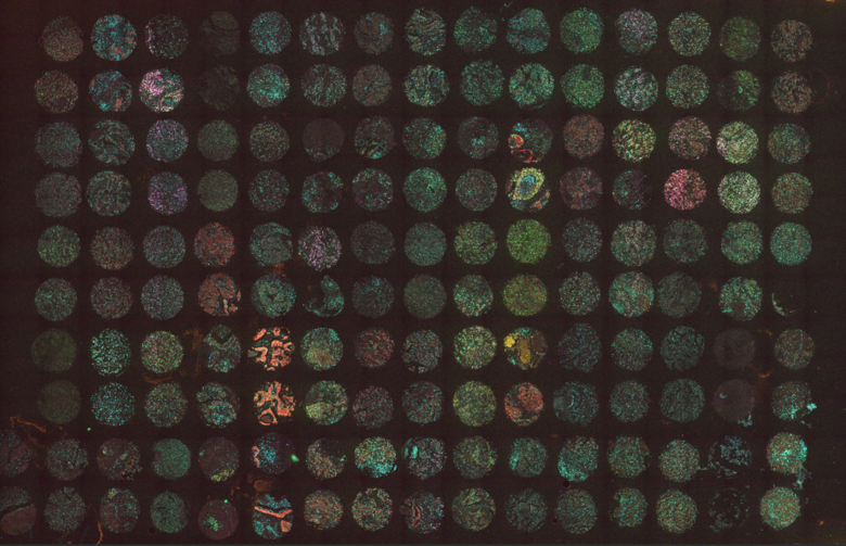
MUSE® 4-plex immunofluorescence on paired FFPE cores (breast cancer)
Cancer research has moved far beyond what can be learned from a single biomarker. Researchers need to understand how malignant cells interact with immune cells, stromal structures, vasculature, and signaling niches within the tumor microenvironment. This spatial context matters because tumors are not uniform masses of identical cells. Rather, tumors are complex ecosystems shaped by heterogeneity, immune infiltration, local inflammation, metabolic stress, tissue architecture, therapy pressure, and, ultimately, therapy response.
For cancer biology, tumor immunology, pathology, and translational oncology labs, this creates a practical technical challenge: how can researchers detect multiple biomarkers in precious FFPE tissue while preserving morphology, maintaining spatial resolution, and generating reliable signal from weak or low-abundance targets?
An ideal workflow should support multiplex staining with user-selected validated antibodies, reduce the species and isotype constraints associated with conventional secondary-antibody approaches, provide signal amplification and a high signal-to-noise ratio, simplify assay setup, and fit smoothly into reproducible research pipelines.
Why the Tumor Microenvironment Requires Spatial Multiplexing
The tumor microenvironment is composed of cancer cells, immune cells, fibroblasts, endothelial cells, extracellular matrix, cytokine gradients, and suppressive or inflammatory niches. A single-marker spatial assay can show where one protein is present, and morphology may sometimes suggest the expressing cell type. However, it cannot reliably resolve co-expression, multi-marker cell states, or spatial relationships among several tumor, immune, and stromal populations in the same tissue section.
For example, a tumor may contain cytotoxic T cells, regulatory T cells, macrophages, dendritic cells, exhausted or checkpoint-associated immune subsets, and tumor cells with different differentiation or activation states. Their biological significance often depends on location. Are T cells excluded from tumor nests? Are macrophages clustered near invasive margins? Are immune checkpoints expressed on tumor cells, immune cells, or both? Are rare low-abundance markers present in spatially restricted regions?
These questions are especially relevant in immuno-oncology, where tumor response or resistance may depend on the organization of immune phenotypes rather than total immune-cell abundance alone. Multiplex immunofluorescence gives researchers the ability to examine several markers in the same tissue section, preserving spatial relationships that are often lost in bulk molecular assays or serial single-marker staining.
The FFPE Challenge in Oncology Research
Formalin-fixed, paraffin-embedded tissue remains one of the most important sample types in oncology research. FFPE samples are widely available, clinically relevant, and often linked to pathology records, treatment histories, or clinical outcomes. However, FFPE tissue also brings well-known technical obstacles that tend to make it more challenging to work with than cryosections, fresh-frozen tissue, or adherent cell samples.
Formalin fixation can mask epitopes, making some biomarkers difficult to detect without optimized antigen retrieval. Tissue age, fixation duration, processing conditions, and storage can all affect antigen quality. FFPE tumor sections can also show variable autofluorescence from fixation-related artifacts, endogenous pigments, red blood cells, necrotic regions, and stromal components. In oncology studies, these challenges are amplified by limited sample availability: small biopsies, rare tumor specimens, and archival cohorts may leave little room for repeated staining attempts or failed optimization rounds.
Robust assay design is therefore critical. Researchers need methods that maximize biological information from each section while minimizing tissue loss, morphology damage, background, and variability introduced by repeated processing steps. Signal amplification cannot replace antibody validation, antigen retrieval optimization, or autofluorescence controls. However, by increasing target-associated signal relative to background, it can make weak epitopes, aged specimens, and background-prone tissue regions easier to interpret.
The Practical Limits of Cyclic Multiplexing
Cyclic staining approaches can expand multiplexing capacity by staining, imaging, stripping, and re-staining the same tissue section. When optimized, these methods can be powerful. They can also be demanding: each stripping or bleaching cycle introduces potential risks, including tissue detachment, morphology damage, epitope degradation, incomplete signal removal, or increased background.
For fragile FFPE sections, small biopsies, or tissues with delicate morphology, repeated cycles can be particularly challenging. The more rounds required, the greater the chance that tissue quality declines before all markers are captured, potentially leading to marker-dependent signal loss or inconsistent detection in later cycles. This can compromise downstream analysis and reduce confidence in interpretation, especially when evaluating weak, heterogeneous, or borderline staining patterns. These challenges can also translate into long optimization timelines and higher assay-development costs. In some cases, substantial optimization effort may still fail to produce a robust panel.
A simultaneous direct or low-cycle multiplex strategy, such as an immunoMUSE® Activate & Amplify 4-plex workflow, can help balance biological insight with practical robustness. Four well-chosen markers can often define key tumor, immune, stromal, or activation states without requiring extensive cyclic processing. This is especially useful in early-stage research, preclinical development, biomarker screening, and translational oncology workflows where reproducibility and turnaround time matter.
Detecting Low-Abundance Biomarkers
Many biologically important cancer and immune markers are not highly expressed. Cytokines, transcription factors, checkpoint molecules, activation markers, phospho-signaling markers, and rare cell-population markers may produce faint staining with standard immunofluorescence. This creates a major challenge for tumor microenvironment research, where important signals may be subtle, tightly localized, or present only in a small subset of cells.
Low signal can be consequential. It can lead to false negatives, underestimation of target expression, and uncertainty in borderline cases. In breast cancer, HER2-low and HER2-ultralow expression states highlight how faint membrane-associated signal can matter. These low-expression categories are increasingly relevant to treatment eligibility and increasingly relevant in translational oncology and diagnostic-development contexts, making accurate detection important for translational research and diagnostic development. In immuno-oncology, heterogeneous or borderline PD-L1 staining can affect how researchers interpret tumor-immune interactions. In both cases, insufficient sensitivity can turn biologically meaningful low expression into apparent absence.
Signal amplification addresses this sensitivity problem by increasing specific target-associated signal relative to background. The point is not simply to make images brighter. The goal is to make weak, spatially restricted, or borderline biomarkers measurable enough to support confident interpretation. A useful amplification workflow should therefore improve signal-to-background while preserving morphology, spatial resolution, and compatibility with multiplex staining.
Managing Autofluorescence and Background
Autofluorescence is a frequent obstacle in FFPE tumor tissue. Endogenous tissue components, fixation artifacts, red blood cells, necrotic regions, collagen-rich stroma, and lipofuscin-like pigments can all contribute to background fluorescence. In tumors, this can be especially problematic because morphology is heterogeneous: necrotic areas, stromal regions, immune infiltrates, and tumor nests may each have different background profiles.
High background reduces fluorescence signal-to-noise ratio and makes weak signals harder to interpret. This is particularly relevant when detecting low-abundance immune markers or subtle biomarker expression. A workflow that improves signal-to-noise ratio can increase confidence that observed staining reflects true biology rather than tissue autofluorescence or nonspecific binding.
Autofluorescence should also be managed through reliable assay design, including appropriate controls such as unstained controls, single-marker controls, no-primary controls, and careful channel selection. Bright and photostable fluorophores should be assigned to weaker targets, while highly expressed markers can often be paired with less intense channels. Imaging settings should also be standardized to avoid artificially amplifying background or saturating true signal.
Panel Flexibility: Using Validated Antibodies
Cancer is a notoriously complex disease, and panel flexibility is required to tackle that complexity. Marker selection depends on the tumor type, model system, treatment mechanism, and biological question. A breast cancer study focused on epithelial identity and proliferation may require a very different panel from a melanoma or lung cancer study examining checkpoint biology, antigen presentation, macrophage localization, or stromal activation.
This is why open antibody compatibility is so valuable in oncology. Multiplex IHC and IF are widely used to define complex immunophenotypes, quantify immune-cell subsets, and assess the spatial arrangement of marker expression in the tumor microenvironment. However, these assays are critically dependent on careful antibody selection, optimization, and validation before they can generate reliable biological data.
Ideally, researchers should not have to compromise on antibody quality to make a panel work. The best panel should be built from the strongest antibodies for each target, not merely from the subset of antibodies that happens to fit a restrictive multiplex format.
An immunoMUSE® Activate & Amplify workflow designed for broad compatibility with user-selected validated antibodies allows researchers to build panels around their own biology rather than relying only on predefined marker sets. As with any immunostaining method, individual antibody clones still require appropriate validation and assay optimization, but researchers can start from antibodies they already trust in their samples and applications.
Practical 4-Plex Panel Concepts in Oncology
A compact 4-plex panel can be highly informative when the markers are chosen around a focused biological question. One study may combine a tumor lineage marker, Ki-67, cleaved caspase-3, and PD-L1 to evaluate proliferation, cell death, and checkpoint-ligand expression in the same tissue section. Another may use CD8, FOXP3, PD-1, and a tumor marker to distinguish cytotoxic and regulatory T-cell populations while assessing checkpoint-associated PD-1 expression in relation to tumor nests. A macrophage-focused panel could include CD68, CD163, HLA-DR, and a stromal or tumor marker to examine myeloid localization, antigen-presentation status, and macrophage activation-associated phenotypes.
The advantage is not simply more markers per slide. It is the ability to connect marker expression to tissue architecture, cell identity, and spatial relationships in the same section. This is particularly important in translational oncology, where confidence in antibody clone performance, staining pattern, and tissue context can determine whether a candidate biomarker is suitable for further validation.
Conclusion
Oncology and tumor microenvironment research increasingly depends on spatial, multiplexed, and tissue-preserving methods. Researchers need to detect low-abundance biomarkers, resolve immune phenotypes, study tumor heterogeneity, and validate biomarkers in FFPE tissue without exhausting precious samples or damaging morphology.
Across cancer biology, tumor immunology, and translational oncology, the shared requirement is clear: flexible multiplex staining, compatibility with validated antibodies, strong signal amplification, high signal-to-noise ratio, preserved spatial resolution, and robust workflows for limited samples.
As cancer research becomes more spatially informed and more personalized, the most useful technologies will be those that help scientists see more biology from less tissue. A practical multiplex immunofluorescence workflow can bridge discovery research and translational validation by making complex tumor ecosystems clearer, more measurable, and more biologically meaningful.