Immunofluorescence is one of the most powerful techniques for visualizing protein localization, cell structure, and biomarker expression. When immunofluorescence works well, it delivers information-rich images that can be strikingly beautiful. When it goes wrong, it can produce high background, weak signal, misleading localization, or complete experimental failure. In many cases, the difference between success and frustration comes down to a handful of avoidable mistakes in sample preparation, antibody setup, and imaging.
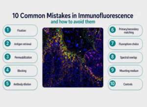
Below are some of the most common pitfalls in immunofluorescence and practical ways to prevent them.
1. Using the Wrong Fixation Method
One of the earliest and most important decisions in immunofluorescence is fixation. This step preserves cell and tissue structure, but not all fixatives behave the same way. A common mistake is assuming one protocol fits every target.
For example, formaldehyde is widely used because it preserves morphology well by crosslinking proteins. However, crosslinking can also mask epitopes, making antibody binding more difficult. In contrast, methanol fixation precipitates proteins and can work well for some cytoskeletal targets, but it may disrupt membrane structures and destroy certain epitopes.
Choosing the wrong fixative can reduce signal, alter localization, or create inconsistent results.
How to avoid it: Always optimize fixation for the antigen and sample type. Start by checking whether your target is known to perform better with formaldehyde or methanol, for example. For membrane proteins and many delicate structures, formaldehyde is often preferred. For some intracellular proteins, especially certain cytoskeletal markers, methanol may work better. If signal is poor, fixation should be one of the first variables you revisit.
2. Skipping or Misapplying Antigen Retrieval and Pretreatment
A frequent mistake in tissue immunofluorescence is neglecting antigen retrieval or using the wrong pretreatment. Crosslinking fixatives, especially formaldehyde-based ones, can conceal epitopes. Without retrieval, the antibody may never gain proper access to the target.
On the other hand, excessive retrieval can damage tissue architecture, increase background, or destroy antigenicity.
How to avoid it: Treat antigen retrieval as a target-specific optimization step, not a default step. Heat-induced antigen retrieval or enzymatic pretreatment may improve staining dramatically, but conditions must be matched to the tissue, fixation conditions, and antigen. If your signal is weak or absent in fixed tissue, test antigen retrieval buffers and conditions systematically rather than assuming the antibody is at fault.
3. Over- or Under-Permeabilizing the Sample
Improper permeabilization is another major source of poor immunofluorescence data. If permeabilization is too mild, antibodies cannot access intracellular targets. If it is too harsh, membranes become overly disrupted, soluble proteins may be lost, and background can rise.
This is especially problematic when working with nuclear or cytoplasmic antigens, where access matters, but preservation matters just as much.
How to avoid it: Choose the permeabilization reagent and concentration according to the localization of your target. Mild detergents may be enough for many intracellular antigens, while stronger treatments can be detrimental. Optimize alongside fixation, since the two steps interact closely. A protocol that works after formaldehyde fixation may not behave the same way after methanol fixation.
4. Inadequate Blocking Leads to Non-Specific Staining
High background and non-specific staining often come from poor blocking. Some researchers rush through this step or use a blocker that is incompatible with the antibody system. The result is diffuse fluorescence, false-positive signal, or staining in unexpected cellular compartments.
Blocking is meant to reduce unwanted interactions between antibodies and the sample. But it cannot compensate for every problem, especially if antibody concentration is too high or species matching is incorrect.
How to avoid it: Use an appropriate blocking reagent for your sample and detection strategy. Serum, protein-based blockers, or specialized commercial buffers can all work, but they should be selected with awareness of the species and assay design. Extend blocking time if background is persistent, and combine blocking optimization with careful antibody titration. Good blocking reduces non-specific binding, but only when the rest of the protocol is also appropriate.
5. Incorrect Antibody Dilution
One of the most common and underestimated errors is incorrect antibody dilution. Too concentrated, and antibodies generate high background, off-target binding, and non-specific staining. Too dilute, and the signal becomes weak or disappears entirely.
There is no universal “best” dilution. An antibody that works beautifully at one concentration in cultured cells may fail completely in tissue sections or 3D samples.
How to avoid it: Run a dilution series for both primary and secondary antibodies. This is especially important when using a new lot, new sample type, or modified fixation protocol. Do not rely blindly on the datasheet. Recommended dilutions are useful starting points, not final answers. Optimization saves time in the long run and produces far more reliable data.
6. Mismatching Primary and Secondary Antibodies
Problems often arise from poor planning around matching primary and secondary antibodies. Secondary reagents must recognize the host species and isotype of the primary antibody. If they do not, signal may be absent, reduced, or misleading.
This issue becomes even more important in multiplex experiments. If multiple primaries come from the same host species or share the same isotype, cross-reactivity will complicate interpretation.
How to avoid it: Confirm isotype compatibility and species reactivity before you begin. Make sure the secondary antibody is designed for the exact host in which the primary was raised. Also consider antigen compatibility when multiplexing. Some antibody pairs may work individually but interfere when combined in a panel. This is especially relevant when using several monoclonals from the same species, or when mixing polyclonals and monoclonals in complex workflows.
Polyclonals can provide strong signal because they recognize multiple epitopes, but they may also increase background. Monoclonals are often more specific, but they may be more sensitive to fixation or epitope masking. Neither is inherently better in all situations; the right choice depends on the target and application.
7. Poor Fluorophore Selection
Not all fluorophores are equally suitable for every experiment. A common mistake is selecting dyes based only on brightness or convenience without considering spectral overlap, instrument configuration, or sample autofluorescence.
Some fluorophores are bright but prone to rapid fading. Others have better photostability and are more suitable for long imaging sessions or repeated scanning. Poor fluorophore choice can also create problems with crosstalk and bleedthrough, especially in multicolor panels. For more information visit Understanding Fluorophores: Choosing the Right Fluorescent Dyes for Your Experiment
How to avoid it: Choose fluorophores based on the full experimental setup. Consider brightness, photostability, expected antigen abundance, and spectral separation. Bright dyes may be reserved for low-abundance targets, while more robust dyes can be used for frequent imaging or archival work. Match fluorophore emission profiles to your microscope’s available excitation lines and detection filters. A strong fluorophore on paper is not useful if your instrument cannot excite or separate it properly.
8. Ignoring Spectral Overlap and Imaging Artifacts
Multicolor immunofluorescence can be visually impressive, but it is also easy to misinterpret. If fluorophore emissions overlap, one channel can appear in another, leading to bleedthrough. This can create false colocalization and distort quantitative analysis. More broadly, crosstalk between channels is a major common source of confusion in poorly designed panels.
Even a well-stained sample can look wrong if the microscope settings are not optimized.
How to avoid it: Design panels with spectral separation in mind. Use fluorophores whose excitation and emission profiles fit your available laser lines and filters with minimal overlap. Acquire single-stained controls to assess bleedthrough and establish compensation strategies where needed. Sequential scanning can also reduce crosstalk in some systems. Never assume that apparent overlap means true biological colocalization until imaging artifacts have been ruled out.
9. Using the Wrong Mounting Medium
The mounting medium is often treated as an afterthought, but it can strongly affect image quality and signal longevity. Some media reduce photobleaching, while others are better for refractive index matching or long-term storage. An incompatible mounting medium can alter fluorescence intensity, increase background, or reduce stability over time.
How to avoid it: Select a mounting medium that matches your fluorophores and imaging goals. Antifade formulations are often valuable, particularly when using less photostable dyes. If samples will be stored before imaging, confirm that the medium preserves fluorescence adequately. A good staining protocol can still be undermined by a poor final mounting choice.
10. Failing to Use Proper Controls
Many immunofluorescence mistakes become obvious only when controls are included. Without controls, it is difficult to distinguish true signal from non-specific staining, autofluorescence, secondary-only artifacts, or fixation-related changes.
How to avoid it: Include no-primary controls, secondary-only controls, and, when appropriate, positive and negative biological controls. For multiplex panels, single-color controls are essential for evaluating crosstalk and bleedthrough. Controls may seem like extra work, but they are often the fastest way to identify the real source of a problem.
Final Thoughts
Immunofluorescence is both a science and a craft. Many failed experiments trace back not to bad antibodies or poor samples, but to avoidable decisions made during preparation and imaging. Errors in fixation, antigen retrieval, pretreatment, permeabilization, blocking, antibody dilution, fluorophore selection, and microscope setup can all compromise results.
The good news is that most of these problems are preventable. By paying close attention to matching primary and secondary antibodies, checking isotype compatibility and antigen compatibility, choosing between polyclonals and monoclonals thoughtfully, minimizing non-specific staining, selecting fluorophores with good photostability, and accounting for laser lines, filters, crosstalk, bleedthrough, and mounting medium, researchers can dramatically improve both signal quality and confidence in interpretation. In immunofluorescence, success rarely comes from shortcuts. It comes from careful optimization, strong controls, and respect for the details that shape every image.