Blog
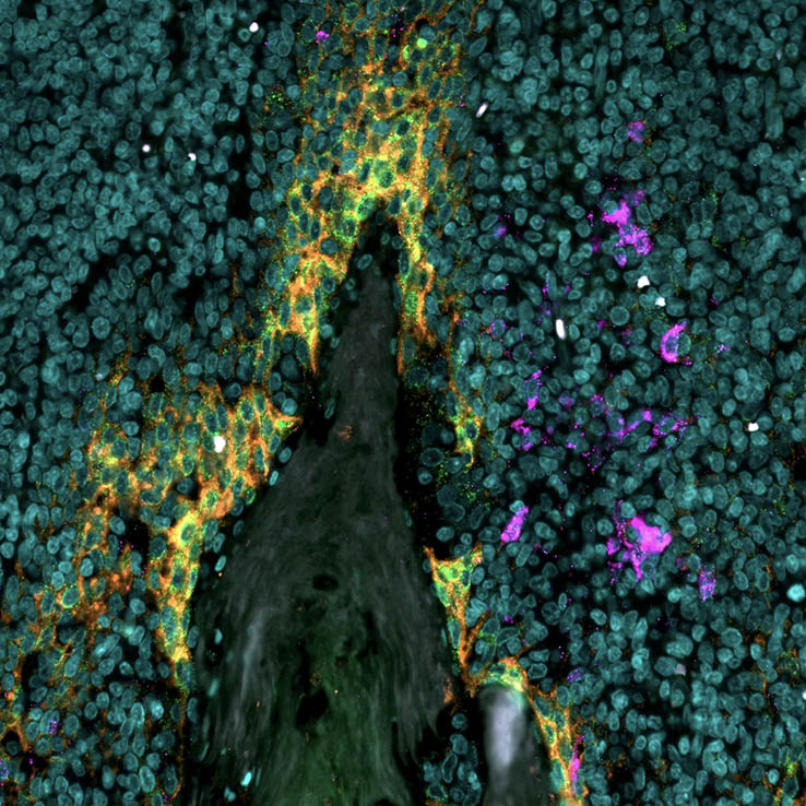
Cancer research has moved far beyond what can be learned from a single biomarker. Researchers need to understand how malignant cells interact with immune cells, stromal structures, vasculature, and signaling niches within the tumor microenvironment. This spatial context matters because tumors are not uniform masses of identical cells. Rather, tumors are complex ecosystems shaped by heterogeneity, immune infiltration, local inflammation, metabolic stress, tissue architecture, therapy pressure, and, ultimately, therapy response.
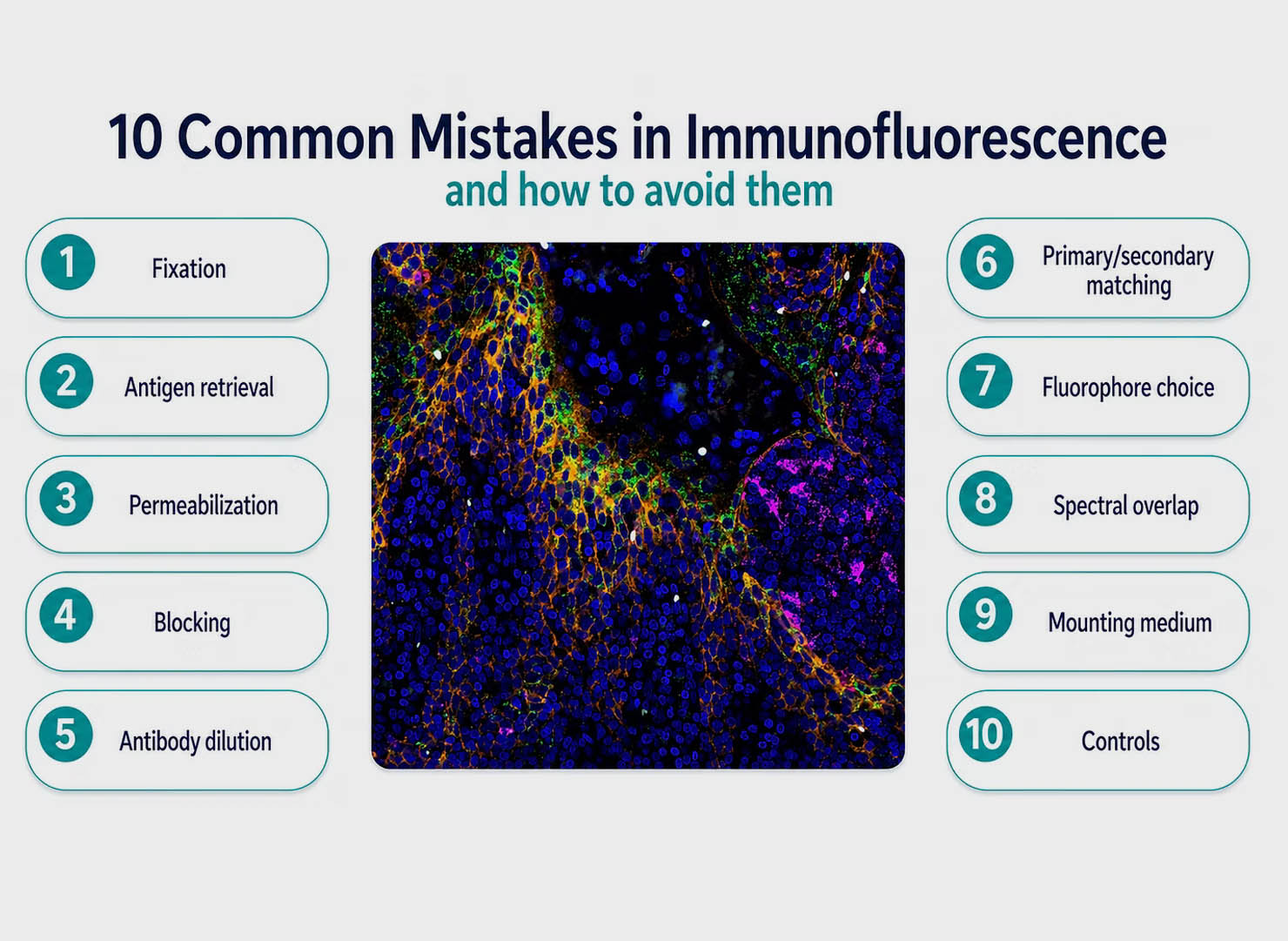
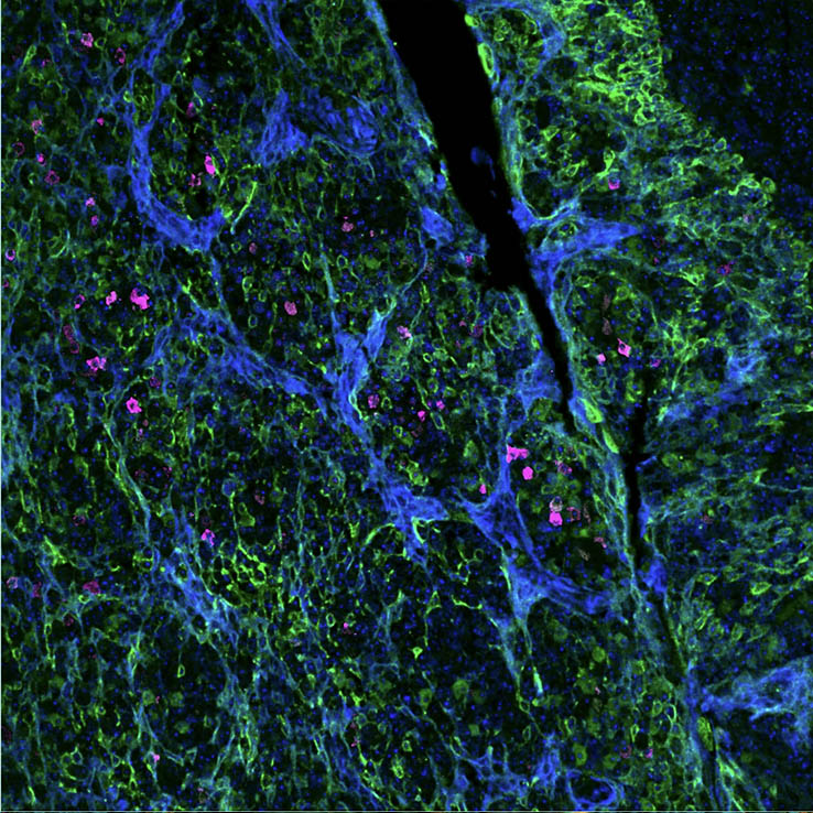
Immunofluorescence is one of the most powerful techniques for visualizing protein localization, cell structure, and biomarker expression. When immunofluorescence works well, it delivers information-rich images that can be strikingly beautiful. When it goes wrong, it can produce high background, weak signal, misleading localization, or complete experimental failure.
Fluorescence-based techniques have become fundamental tools in modern biological and biomedical research. From visualizing cellular structures to tracking molecular interactions, fluorescence imaging enables scientists to observe biological processes with remarkable precision.
Immunofluorescence microscopy (IF microscopy) is a cornerstone technique in modern life sciences. By combining the specificity of antibodies with the sensitivity and spatial resolution of fluorescence microscopy techniques, researchers can visualize the precise localization of biomolecules in cells and tissues. Whether you are a novice exploring fluorescent labeling or a seasoned researcher aiming for multiplexed imaging, understanding direct immunofluorescence, indirect immunofluorescence, and the nuances of immunofluorescence staining is essential.
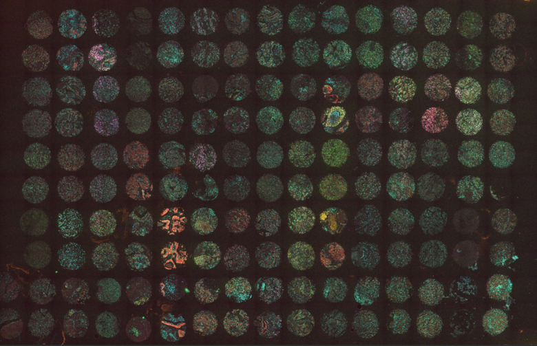
Uncovering how biology underlies health and disease requires more than measuring individual markers—it requires knowing where these markers are located and how cells interact within tissues. Spatial biology techniques preserve tissue architecture to reveal complex molecular and cellular interactions. Combined with multiplex imaging and spatial omics, researchers can study multiple biomarkers simultaneously, enabling deep insights into tissue organization, immune landscapes, and tumor microenvironments.